
America’s #1 killer has new foes: FIU tackles heart disease with early detection, robotic surgery and next generation cardiac care
A hypnotic rhythm underlies your existence. It began before you were born. It continues, right this very moment, without a single conscious thought on your part. It will be with you until the end. Lay a hand to your chest. Feel it. Ba-bum. Ba-bum. Ba-bum.
The heart is the muscle that works harder than any other. It contracts as if being squeezed then relaxes, over and over and over again, around 100,000 times a day. That’s roughly three billion times in an average lifespan. With each beat, the heart valves take turns opening and snapping shut in a well-orchestrated dance to keep oxygen-rich blood flowing through the branching network of arteries and veins stretching from the top of your head down to your toes.
The heart’s job is relatively straightforward, even simple. The heart itself is remarkably complex. Its many marvels and enduring mysteries have been an endless source of fascination for millennia. Aristotle considered it the most important organ. French philosopher René Descartes saw it as a sort of engine. Leonardo da Vinci filled countless pages of his famous notebooks with intricately detailed ink-and-quill anatomical studies, in many cases hypothesizing correctly about its function. Beneath one such drawing, he wrote:
“How in words can you describe this heart without filling a whole book?”
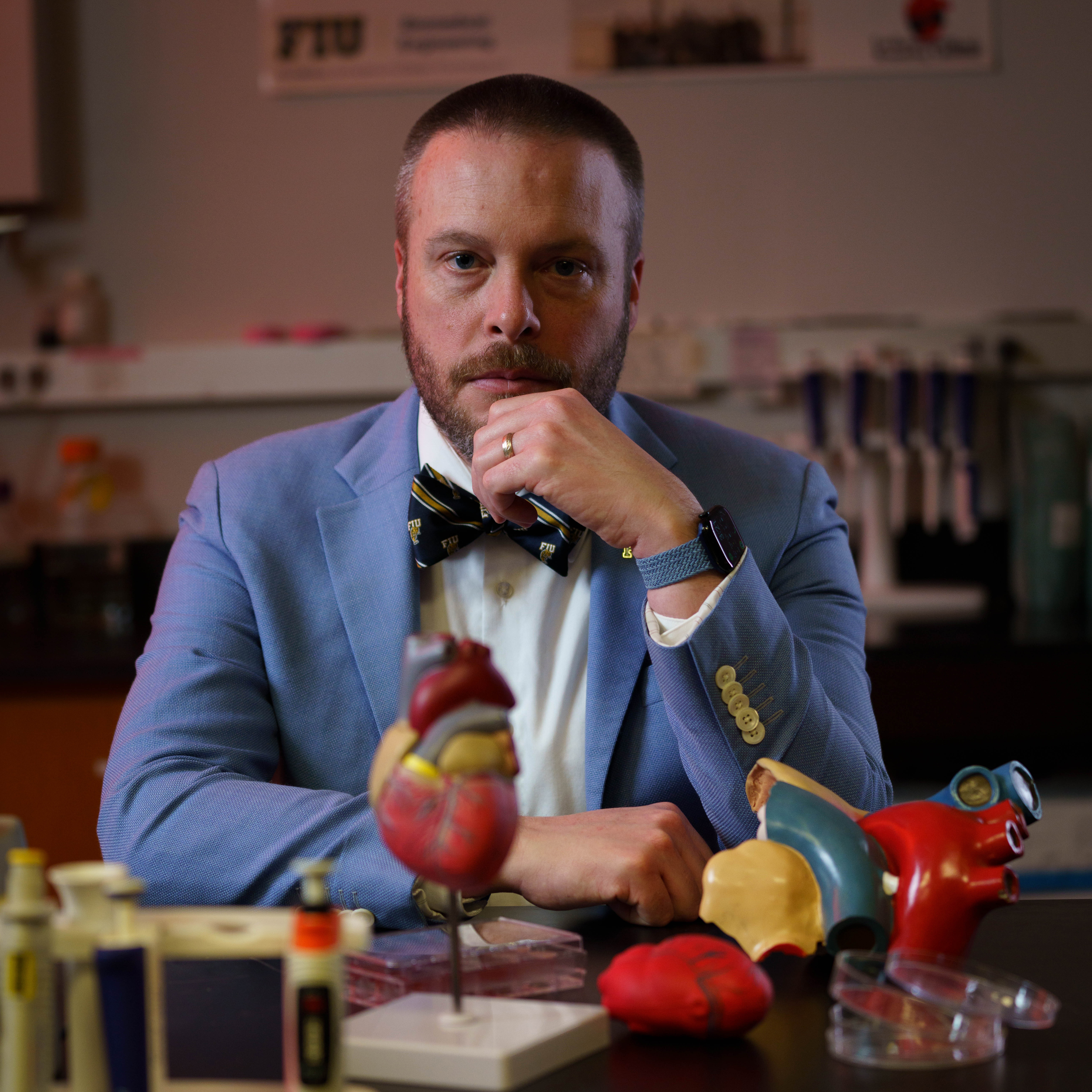
Indeed, how? Even today, researchers like Joshua Hutcheson — a biomedical engineer and Fellow of the American Heart Association who has studied the heart’s intricate mechanisms for the better part of a decade — can attest to the fact that understanding the heart remains an awe-inspiring journey into the unknown. In lectures, he shows students Leonardo’s drawings. On the wall of his Cardiovascular Matrix Remodeling Lab at FIU hangs a quote from Descartes. Both are nods to the origins of modern physiology as much as reminders to embrace the kind of curiosity that drove the great thinkers of history.
Asking big, bold and daring universal questions — the why and how of life — is almost a prerequisite when it comes to the heart. “That’s how you get to a place where you discover something completely new,” says Hutcheson.
Here’s one: Why does the heart work flawlessly throughout an entire lifetime for some and not for others?
Cardiovascular disease — an umbrella term encompassing a number of conditions, including coronary heart disease, heart failure, arrhythmias (i.e. atrial fibrillation), vascular disease, congenital heart defects and more — has been the nation’s leading cause of death for over a century.
The good news: Death rates have steadily declined over the decades, thanks in part to widely available medications that control blood pressure and artery-clogging cholesterol, as well as successful public health campaigns to stop smoking. The not-so-good news: The downward trend is stalling. An aging population — expected to drive a surge in cardiovascular disease burden — is on a collision course with a growing number of younger adults with risk factors, such as uncontrolled blood pressure, diabetes, and obesity.
The American Heart Association estimates at least six in 10 U.S. adults (more than 184 million people) will have some type of cardiovascular disease by 2050.
At FIU and beyond, researchers like Hutcheson as well as physician-scientists and cardiac surgeons focus their life’s work around addressing this problem. In their labs and the clinic, they are leading major technological and life-saving therapeutic advances to gain a deeper understanding of how cardiovascular disease happens and transform how it is prevented, detected and treated.
Silent beginnings
There’s a lot about cardiovascular disease that’s vexing. Perhaps most frustrating is how it moves in stealth. “By the time symptoms appear, the heart has been irreparably damaged,” explains Hutcheson.
Atherosclerosis, the buildup of plaque in the arteries, is a major culprit of disease and slow-ticking time bomb. In fact, it can start in childhood.

Plaque — made up of cholesterol, inflammatory cells and other material — can collect inside artery walls. Over time, these fatty deposits narrow the artery, reducing blood flow. Problems happen when plaque becomes unstable and ruptures. Blood clots form at the rupture site. Those clots can block blood flow. If it happens in the heart, it causes a heart attack.
But rewind: How does this happen in the first place? Hutcheson initially set out searching for the earliest possible mechanisms behind this mineralization as a postdoctoral researcher at Harvard.
After joining the lab of Elena Aikawa — a leading expert in cardiovascular calcification — Hutcheson created a series of models that mimicked vascular tissue and arterial cells. Up close and in real time, he watched as nanosized membrane sacs resembling bubbles, called extracellular vesicles, began to bleb off the cells. One by one, they gathered in clusters. Then things got really interesting: They started “capturing” calcium and phosphate. The vesicles were not only major mediators of calcification. They were the instigators.
With funding from the NIH and Florida Heart Research Foundation, Hutcheson continues to lead far-ranging investigations into these vesicles to find ways to detect or disrupt this process. Cell culture experiments in his lab have had encouraging results: Epidermal growth factor receptor inhibitors, commercially available and FDA-approved for cancer treatment, successfully stopped the vesicles and prevented plaque formation.
Another project is probing the calcification paradox or why it is that the more abnormal calcification present in arteries, the less calcification and mineral development in the bone. Hutcheson and FIU colleagues — including chemist Francisco Fernandez-Lima, biophysicist Prem Chapagain and physicist Jin He — compare how vesicles form and function in bone cells versus artery cells. Early evidence suggests there’s some crosstalk between the two cell types, raising the possibility of coaxing calcium and phosphate from arteries back into bone.
Would it be possible to make plaque completely disappear? Cholesterol-lowering drugs and lifestyle changes, like losing weight and quitting smoking, typically slow the formation of new plaque but don’t get rid of old buildup. Hutcheson’s research group, though, has shown activating a hormone called relaxin could be a step in the right direction. Preclinical mouse models and human cells in vitro demonstrated a small molecule — developed in collaboration with investigators from the National Center for Advancing Translational Sciences — significantly reduced and reversed late-stage vascular calcification.
“This is the dream,” Hutcheson says. “To find a way to reverse the pathology and return patients to a normal baseline.”
Innovating new heart valve solutions
The arteries are, of course, one place plaques appear. The valves another.
Daniel Chaparro spent his entire Ph.D. journey in Hutcheson’s lab studying these little one-way doors of the heart.
“Every time the valves close, I always say it’s like they are clapping for you,” he laughs. “They’re saying ‘Yea! You’re alive!’”
The ‘clapping’ Chaparro refers to is actually audible through a stethoscope: Lub-dub. When the heart contracts, valves between the upper and lower chambers of the heart snap shut (lub). As the heart relaxes, the other two valves that lead out of the heart close (dub) allowing the heart to refill with blood.
If valves don’t open properly, blood trickles through like water when you press a thumb against a hose. When valves fail to close correctly, the opposite happens: blood leaks backward into the heart, leading to congestive heart failure. Both problems can change the sounds of the heart.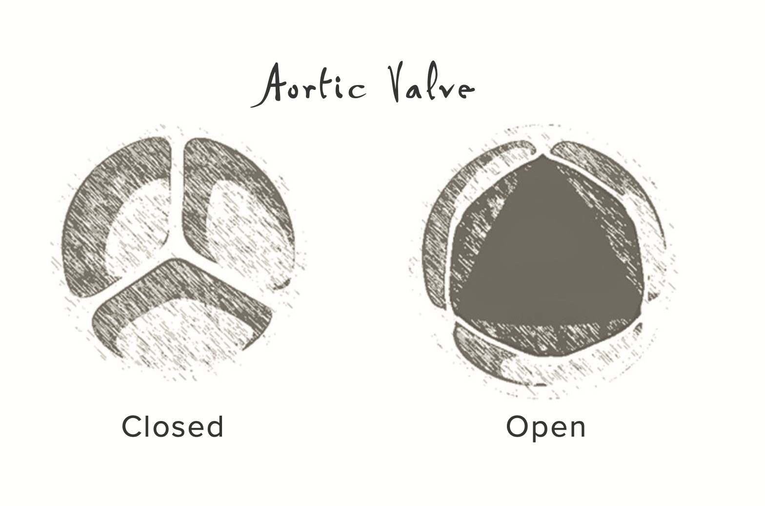
On a computer screen, Chaparro pulls up a magnified image of an aortic valve. It’s made up of three separate flaps, called leaflets. Curiously, calcification doesn’t collect on each leaflet equally. One flap often fares far worse than the others. With a Florida Heart Research Foundation doctoral student grant, Chaparro used computational modeling and experimental planning to pinpoint the possible cell types responsible for these asymmetrical calcification patterns. It was a massive undertaking.
But he’s not quite finished with valves yet. Chaparro is now senior research engineer in the lab of physician-scientist Dr. David Kalfa, a renowned expert in complex neonatal cardiac surgery and advanced pediatric valve repair.
“The times that there have been huge leaps forward, in terms of how we treat or diagnose diseases, is when a surgeon or clinician and engineer get together to find solutions,” Chaparro says. He mentions Dr. Albert Starr and hydraulic engineer Lowell Edwards whose pioneering collaboration produced the world’s first successful artificial heart valve.
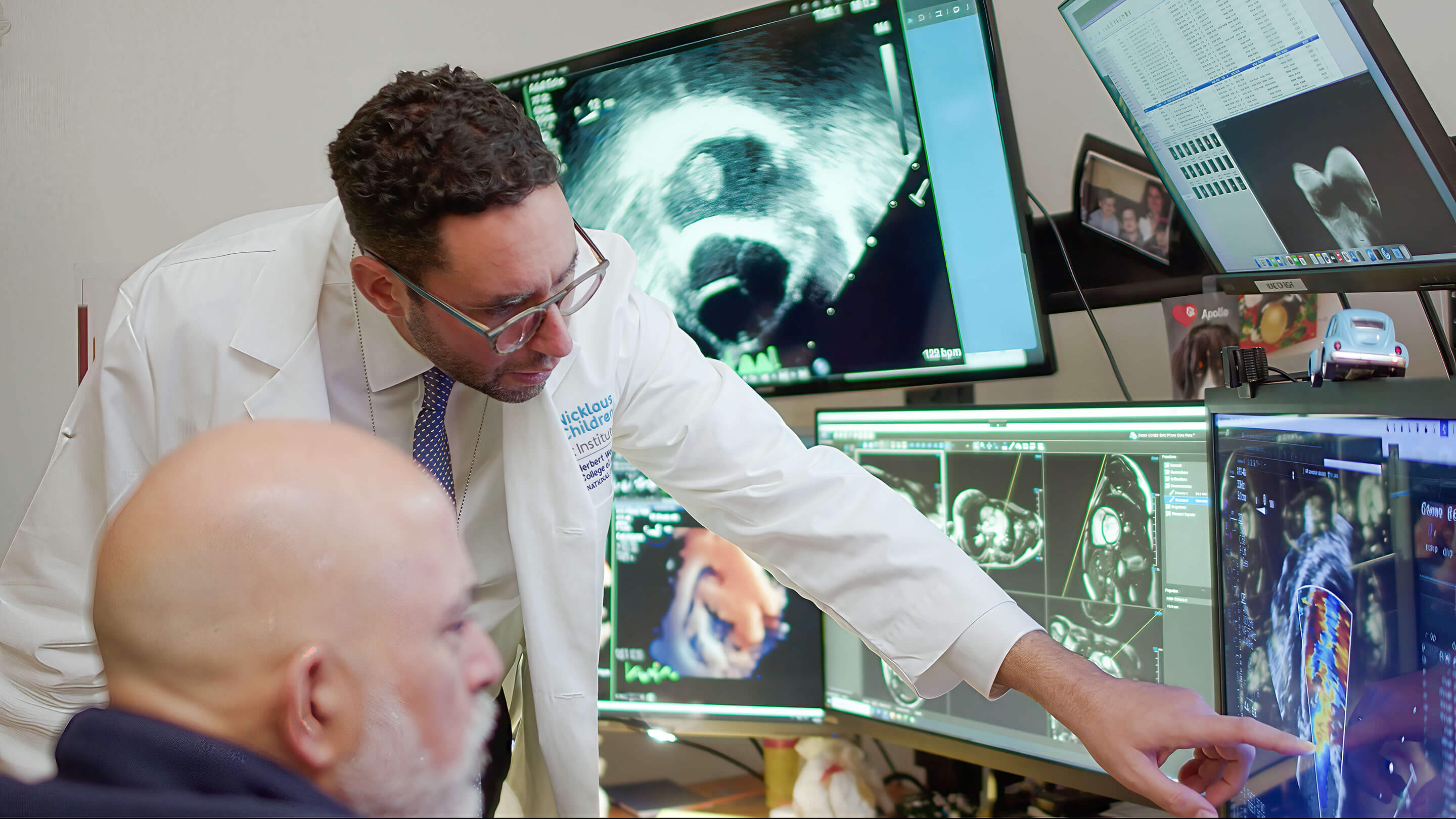
Kalfa’s team has a similar, no less ambitious goal: Find innovative solutions for the estimated 40,000 children born each year with congenital heart defects.
“We still don’t have many good options in terms of heart valve replacement for children with congenital heart defects,” says Kalfa, chief of cardiovascular surgery and co-director of the Nicklaus Children’s Heart Institute, as well as professor in FIU’s departments of pediatrics and surgical sciences. Consider: A newborn heart is around the size of a large strawberry. “The options we do have either aren’t small enough or can’t grow with the baby or child. Each time we need to replace it, that’s another open heart surgery.”
Living allogenic valve transplantation (also called partial heart transplants) has emerged as a promising alternative. Because even if heart muscle is damaged or diseased, the valves can be in good working condition.
In 2023, Kalfa performed the world’s first domino infant partial heart transplant while at New York-Presbyterian Morgan Stanley Children’s Hospital: An 8-month-old received a full heart transplant, and the healthy valves from her old heart were donated to a 2-month-old baby. A year later, Kalfa did a split-root domino partial heart transplant that saved three children in 24 hours. It was an emotional experience, one he says will stay with him forever.
As with many things in life, logistics and timing have to align for partial heart transplants to be a success. That’s why Kalfa’s team is on a mission to extend the extracorporeal time of this precious tissue — and give it an extended shelf life, banking it much like blood is banked, until there is a child who needs it.

With an NIH R01 grant, they will be fine-tuning a unique time-buying approach. Here’s how it works: Tissue is stored in a custom-built bioreactor that closely replicates conditions inside the body, providing pulsing stimulation as a stand-in for a heartbeat. A special solution — patented by Kalfa’s team and proven to keep tissue alive for at least seven weeks — is added to ‘feed’ the tissue and supply essential nutrients.
“This is definitely a new, exciting frontier,” Kalfa says. “It could increase availability and the chance of having the right valve for the right patient at the right time — transforming the lives of children in South Florida, across the country and the world.”
Hearing heart disease
What else could prove life changing for patients? Earlier diagnosis
In addition to looking at the underlying mechanisms of disease, Hutcheson is also developing low-cost, accessible diagnostic tools. He didn’t have to look far for inspiration. He found it close to home. Hutcheson’s wife is a classically trained opera singer. Healthy vocal cords stretch and contract to produce different notes. Damage or injury to these thin folds of tissue can drastically alter the rich resonance and sound of a singer’s voice.
Wait a second, Hutcheson thought, the heart isn’t all that different. When valve disease or calcification occurs, the lub-dub sounds change. Other sounds creep in. Clicks. Whooshes. Extra beats. Could it be possible to train an algorithm to detect the earliest acoustic signatures disease leave behind, even those too subtle for the human ear to hear?
Hutcheson shared the idea with Valentina Dargam, one of his graduate students at the time. The undertaking would be ambitious, a sort of DIY project. For starters, training an algorithm requires data. Lots of it. Dargam would need to collect thousands of heart sounds with a digital stethoscope, analyze the acoustic signals and validate the pre-clinical data against image scans of calcium build-up in the heart. Other people might have hesitated. Dargam didn’t.
“I honestly don’t know how I did everything,” she admits. “But I do know I need to have a vision of ‘Okay, how will this help people one day?’ That drives me.”
Between juggling the demands of the actual research, Dargam signed up for a few commercialization courses. She entered and won several pitching competitions. In 2020, preparing to apply to NSF’s prestigious Innovation Corps (I-Corps) program, she took part in the regional I-Corps training program, interviewing 45 cardiologists and clinicians in the span of five weeks. Their responses were enthusiastic and feedback invaluable. Dargam and Hutcheson were then accepted into the national I-Corps and spent eight weeks ensuring the technology meets future patients’ needs.
Today, the AI-based diagnostic algorithm that Dargam trained is 95% accurate in classifying healthy heart sounds and nearly 85% accurate in differentiating between types of heart disease. It also picks up on disease before cardiac murmurs or structural changes appear. Clinical trials are on the horizon. Dargam and Hutcheson will work with Dr. Tom C. Nguyen — internationally recognized minimally invasive heart surgeon, chief medical executive of Baptist Health Heart & Vascular Care and Barry T. Katzen Endowed Chair at Miami Cardiac & Vascular Institute — to test the technology in the clinic.
“This type of work is critical and the only way to advance medicine,” Nguyen says. “Physicians interact with patients on a daily basis and have a deep understanding of the clinical side of medicine. The team at FIU has a very strong foundation of cardiovascular research. Bringing the two together is a perfect marriage where we all complement each other.”
Robot in the OR
Relevant research that addresses real-world medical needs is at the heart of FIU’s strategic partnerships with Nicklaus Children’s Health System and Baptist Health South Florida. These collaborations have attracted world-renowned talent, including Dr. Makoto Hashimoto, an internationally acclaimed cardiac surgeon pioneering new approaches to robotic-assisted cardiac procedures.
Traditional open-heart surgery requires a six- to eight-inch incision down the center of the chest. The breastbone is cut and ribcage spread open. Recovery can be slow. Hospital stays long. Serious life-threatening complications like infection can occur.
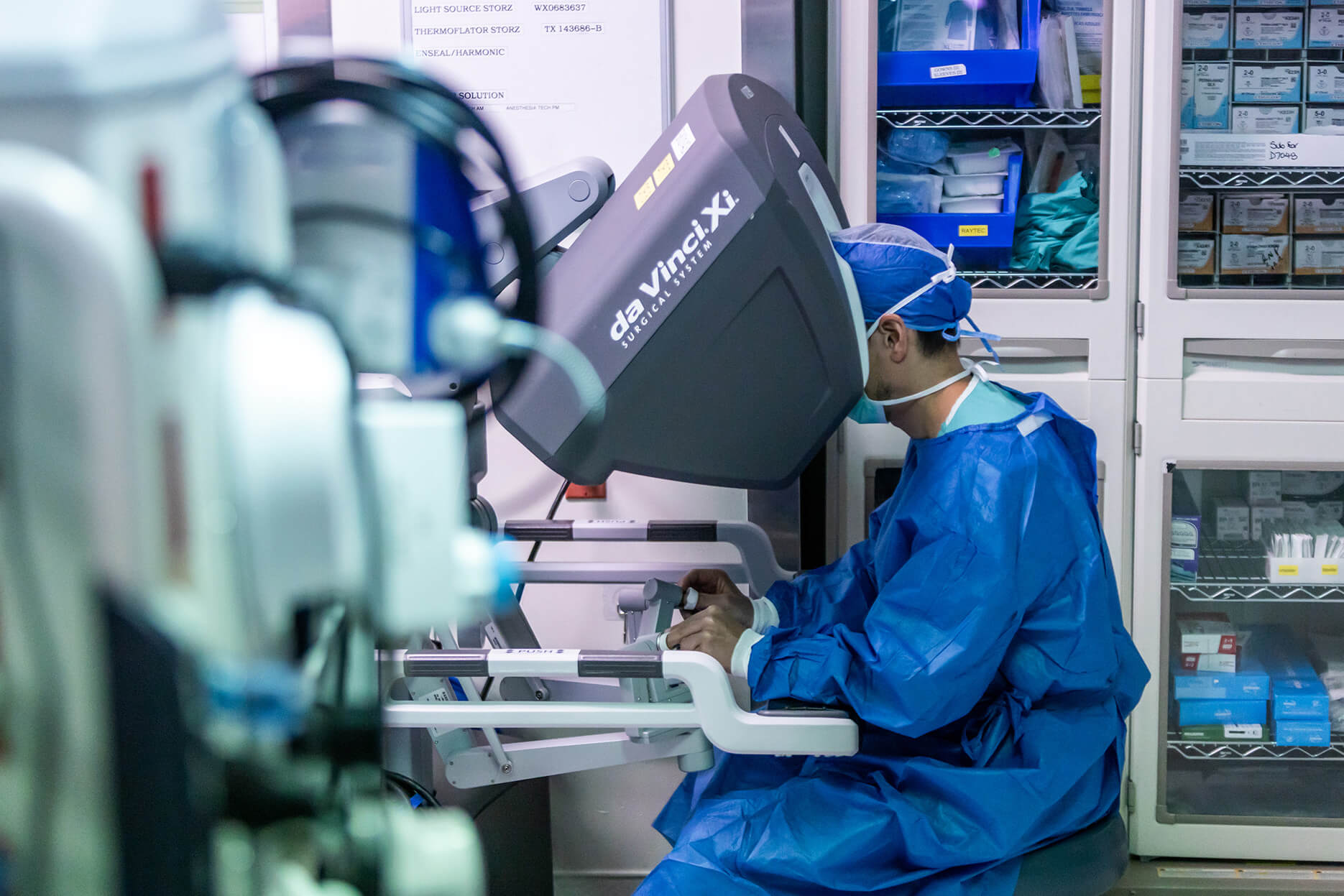
Peek behind the scenes of a robotic-assisted procedure and the scene is dramatically different. Only a few small incisions are made. No bone is cut. A coordinated surgical team of nurses, technicians and an anesthesiologist remain beside the patient’s bedside. Across the room, Hashimoto stays seated at the console connected to a robotic system named after Leonardo da Vinci. He hardly moves, save for the slightest flutter of his fingers on the controller. Every subtle move of his hands is instantly captured, scaled for precision and mirrored by the robotic arms holding the micro-sized instruments and a 3D high-definition camera inside the patient’s chest and heart.
“The robot becomes part of my body during surgery,” says Hashimoto, FIU professor and director of robotic cardiac surgery who treats patients at Baptist Health Miami Cardiac & Vascular Institute. “It feels like an extension of myself.”
Because robotic surgery has many advantages — less pain, minimal scarring, faster recovery — it’s becoming increasingly popular with patients. But widespread adoption is lagging behind other specialties. Few cardiac surgeons perform more than 20 robotic cases a year. Hashimoto, however, performs hundreds. With an ever-growing volume of cases, he’s leading Florida’s busiest and fastest-growing robotic cardiac surgery program.
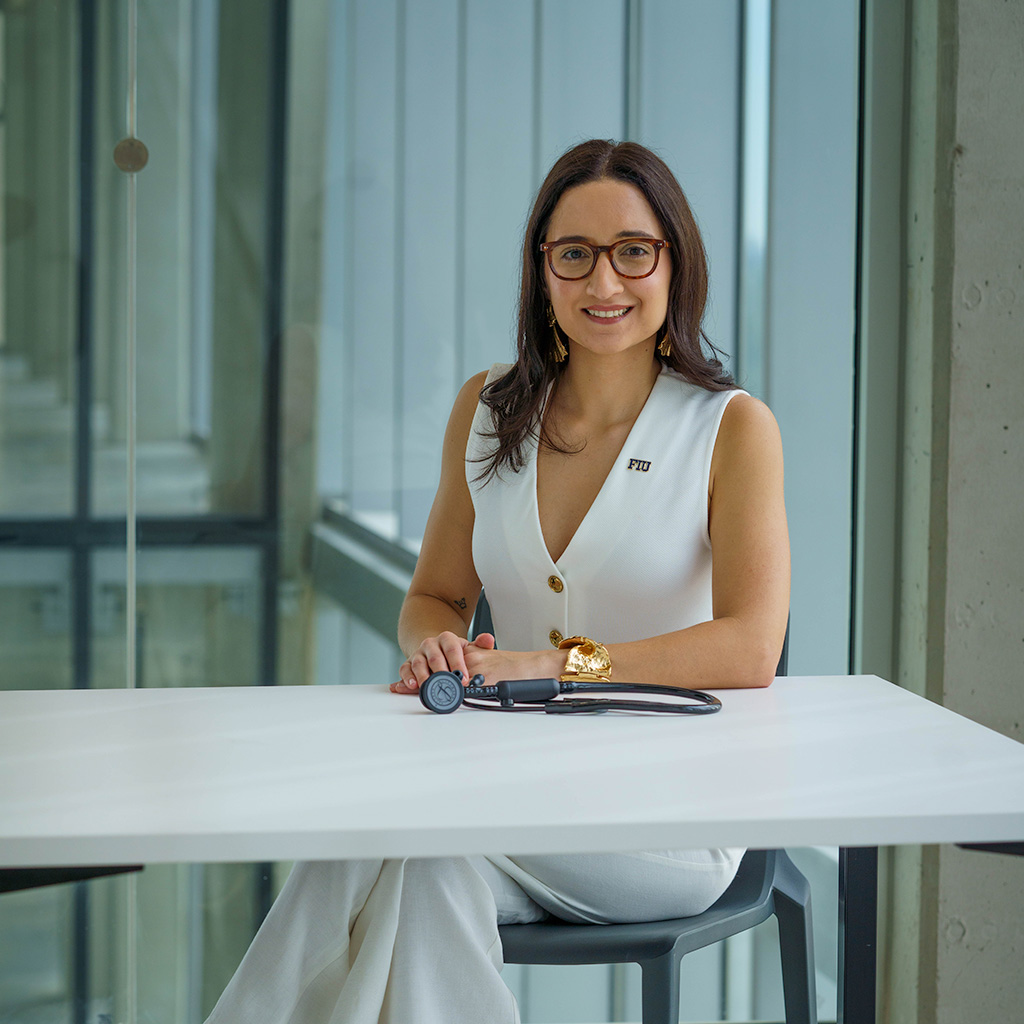
Every surgery Hashimoto completes saves another life. It also helps move the field forward by providing much-needed evidence that demonstrates novel robotic techniques can be applied safely and effectively to various conditions that once required more invasive procedures. FIU medical student Natasha Mazinani, one of Hashimoto’s mentees, is helping advance this important work.
“My grandfather had open heart surgery, so I always thought any heart surgery meant a big scar down your chest,” says Mazinani. “It’s honestly mind blowing to see what Dr. Hashimoto does for his patients. There are no words.”
Mazinani has helped Hashimoto with case studies on complex conditions. One detailed how Hashimoto repaired a heart defect involving both a hole between heart chambers and a leaking valve (known as an atrioventricular septal defect with severe mitral regurgitation). Another retrospective study compares his first 100 coronary artery bypass graft surgeries to his last 100 to explore the learning curve with this type of procedure.
“This research is essential because it’s how we get information out there for other surgeons to learn how to do it and bring these techniques to more patients,” Mazinani says. “I am so grateful to play a small role in advancing what we know about robotic procedures, because it’s a whole new realm. It makes me really excited as a future physician about what advancements are yet to come.”
New era of cardiac research
All this work at FIU continues, following a steady, consistent rhythm of its own.
Hashimoto is planning to expand his scope of research with scientists across the university. Nguyen was recently named chair of the newly formed Department of Cardiovascular Sciences in the Herbert Wertheim College of Medicine. Kalfa wants to establish a congenital heart defect research institute that spans basic science, translational research and clinical trials. Hutcheson, meanwhile, has taken on a new role: inaugural director of the FIU-Florida Heart Research Foundation Center for Innovation in Cardiovascular Health.
Established with $11.7 million from longtime supporter, the Florida Heart Research Foundation, the center is an investment in ongoing multidisciplinary collaborations.
The initial group of center-affiliated researchers will come from across the university. Among them is Dargam. She’s launching her own lab with support from a junior faculty grant and will continue the digital heart sounds project while also focusing on a new line of inquiry: how exposure to heavy metals such as lead and cadmium (environmental toxicants found in various consumer products) contributes to the progression of heart disease.
“It’s a little scary, but also so exciting when you think about the fact I am going to be exploring an entirely new field,” she says. “But I know I can count on everyone who is part of the center. That’s why collaboration is so important. It’s how you advance really cool, important research.”
The center’s official home will, fittingly, be in FIU’s new Innovation Complex. Hutcheson cannot wait to see everyone together — each person essential, each bringing a different expertise and fresh curiosity, each supporting the same purpose.
“When you break down barriers and get a bunch of smart people together to ask a bunch of those fundamental questions, that’s when you start solving big problems,” Hutcheson says. “This is the right place and the right time and the right group of people. Our community and our country need this.”